PET (Positron Emission Tomography) Scans
PET (Positron Emission Tomography) Scans

Positron Emission Tomography (PET) scans are a type of medical imaging that can show how your body's tissues and organs are working, unlike other scans that mainly show structure. Essentially, it allows doctors to see how your body is functioning at a cellular level by tracking a radioactive tracer injected into your body. This can help diagnose a variety of conditions, including cancer, heart problems, and brain disorders.
In short, think of a PET scan as a way to see how your body is using energy and how well different parts are functioning, potentially revealing problems before they become obvious. which can help doctors diagnose and monitor a variety of conditions.
- Radioactive tracers: Imagine a tiny, safe amount of a special substance called a radiotracer is injected into your bloodstream, usually through a vein in your arm. This radiotracer is often a slightly altered sugar (like a radioactive form of glucose called FDG), because many cells in your body, especially active ones like cancer cells or brain cells, use sugar for energy.
- Targeting specific areas: The radiotracer travels through your bloodstream and collects in areas where there's a lot of cell activity, whether it's normal activity or activity related to a disease. For example, cancer cells are often very active and use a lot of sugar, so the FDG tracer will gather in those areas. Other types of PET radiopharmaceuticals are attracted to certain sites on certain cells.
- Imaging the body: You'll lie down on a comfortable table that slides into the PET scanner, which looks like a large doughnut. The scanner detects the tiny amounts of radiation emitted by the radiotracer as it decays in your body.
- Creating images: A computer analyzes the radiation detected by the scanner and creates detailed images of your organs and tissues. These images show how well your organs are functioning, based on how much of the tracer has collected in different areas. Areas with high activity (where the tracer has collected more) appear as bright spots on the images, which can help doctors identify potential problems, like tumors.
Finding and Monitoring Cancer:
- Diagnosing cancer: PET scans can help doctors determine if a tumor is cancerous (malignant) or not (benign). Cancer cells often use more energy, like sugar, than normal cells, and this activity shows up on the scan.
- Staging cancer: If you've been diagnosed with cancer, a PET scan can help determine how far it has spread in your body (staging). This is crucial for planning the most effective treatment.
- Checking if treatment is working: Doctors can use PET scans throughout your treatment to see if the cancer is responding (shrinking or staying the same). This helps them adjust treatment plans as needed.
- Detecting recurrence: After treatment, a PET scan can help check if the cancer has returned.
Evaluating Heart Health:
- Assessing blood flow: PET scans can show areas of reduced blood flow to the heart muscle.
- Evaluating heart damage: This can help determine the effects of a heart attack or identify areas that could benefit from procedures like bypass surgery or angioplasty.
Investigating Brain Disorders:
- Assessing brain function: PET scans can evaluate how different parts of your brain are functioning.
- Diagnosing conditions like: Alzheimer's disease, epilepsy, and stroke.
- Locating surgical sites: In some cases, PET scans can help doctors identify the precise area for brain surgery.
Early Detection:
- Finding problems before they become noticeable: Unlike some other scans that just show what organs look like, a PET scan looks at how your cells are working.
- Spotting trouble early: If something is wrong, like a fast-growing tumor, the cells in that area will be working overtime and will show up as bright spots on the PET scan. This helps doctors catch diseases early, sometimes even before you feel symptoms.
Seeing How Your Organs Function:
- More than just structure: PET scans don't just show the structure of your organs, but also how they're metabolizing (how they're using energy, like sugar).
- Checking your heart: For example, a PET scan can show how blood is flowing to your heart muscle, helping doctors see if there are any issues with blood supply.
- Exploring your brain: It can also show how different parts of your brain are functioning, which is useful for diagnosing and managing conditions like Alzheimer's disease or epilepsy.
Tracking Disease and Treatment:
- Seeing if cancer is spreading: PET scans can help determine if cancer has spread from its original location to other parts of your body.
- Monitoring treatment effectiveness: Doctors can use PET scans to see how well treatments like chemotherapy are working and adjust the plan if needed.
- Finding if cancer has returned: PET scans can also help detect if cancer has come back after treatment.
Radiation Exposure:
- Small amount of radiation: You are exposed to a small amount of radiation from the tracer and from the CT during the scan.
- Low risk of harm: This dose is very low, comparable to a few years of natural background radiation exposure.
- Benefits usually outweigh the risks: The information gained from a PET scan to diagnose or monitor serious health conditions generally outweighs the minimal risks associated with this small dose of radiation.
Allergic Reactions:
- Rare, but possible: Some people may experience an allergic reaction to the radioactive tracer, although this is uncommon.
- Symptoms: Reactions are typically mild, such as itching, hives, or difficulty breathing.
- Severe reactions are rare: Very rarely, a severe, life-threatening allergic reaction called anaphylaxis can occur.
- Inform your doctor: If you have known allergies to medications, contrast dyes, iodine, or latex, be sure to inform your healthcare provider.
Pregnancy and Breastfeeding:
- PET scans generally avoided during pregnancy: If you are pregnant or think you might be, a PET scan is usually not recommended unless absolutely necessary, as the radiation may harm the developing baby.
- Special considerations for breastfeeding mothers: If you are breastfeeding, you may need to temporarily pause breastfeeding after a PET scan, as the tracer can pass into breast milk. Your doctor and technologist will provide specific guidance based on the tracer used.
Discomfort or Pain:
- Lying still: Having to lie still on the scanning table for a prolonged period might cause some discomfort, particularly if you have a recent injury or have undergone surgery.
- Injection site reactions: A slight prick or cold sensation may be felt during the tracer injection. Rarely, you might experience pain, redness, or swelling at the injection site afterward.
Interference with scan accuracy:
- Certain conditions can affect results: High blood sugar levels in people with diabetes, caffeine, alcohol, or tobacco consumed within 24 hours of the scan, and excessive anxiety can all potentially affect the accuracy of the PET scan results.
- Medications: Certain medications, such as insulin and sedatives, can also affect the scan.
- Neurological or psychiatric conditions: Conditions that prevent someone from lying still during the scan can also interfere with the results.
Cardiac PET Scans
A Cardiac Positron Emission Tomography (PET) Scan is a noninvasive nuclear imaging test that uses small amounts of radioactive tracers to create detailed 3D images of your heart. Unlike standard CT or MRI scans that focus on structure, a PET scan reveals how your heart tissues and organs are functioning at a cellular level.
Doctors use these scans primarily to check blood flow, diagnose coronary artery disease (CAD), and evaluate heart muscle damage after a heart attack.
A cardiac PET scan works through a process called "annihilation" to map your heart's function. It uses radioactive tracers that release positrons (tiny particles) that collide with electrons in your body, creating energy that a scanner detects and turns into 3D images.
Tracer Injection & Tracking
- Administration: A technologist injects a radioactive tracer (such as Flurpiridaz F-18) into your bloodstream through an IV.
Cellular Absorption
Heart cells "absorb" these tracers based on their metabolic activity and blood flow. Areas with healthy blood flow will "light up" brightly, while damaged or blocked areas will appear dim or dark.
The Science of the Scan (Positron Emission)
- Positron Release: As the tracer breaks down, it emits positrons.
- Annihilation: These positrons quickly collide with nearby electrons, causing both to be destroyed (annihilated).
- Photon Detection: This collision releases two gamma-ray photons that travel in exactly opposite directions. The donut-shaped PET scanner contains rings of detectors that catch these pairs of photons simultaneously.
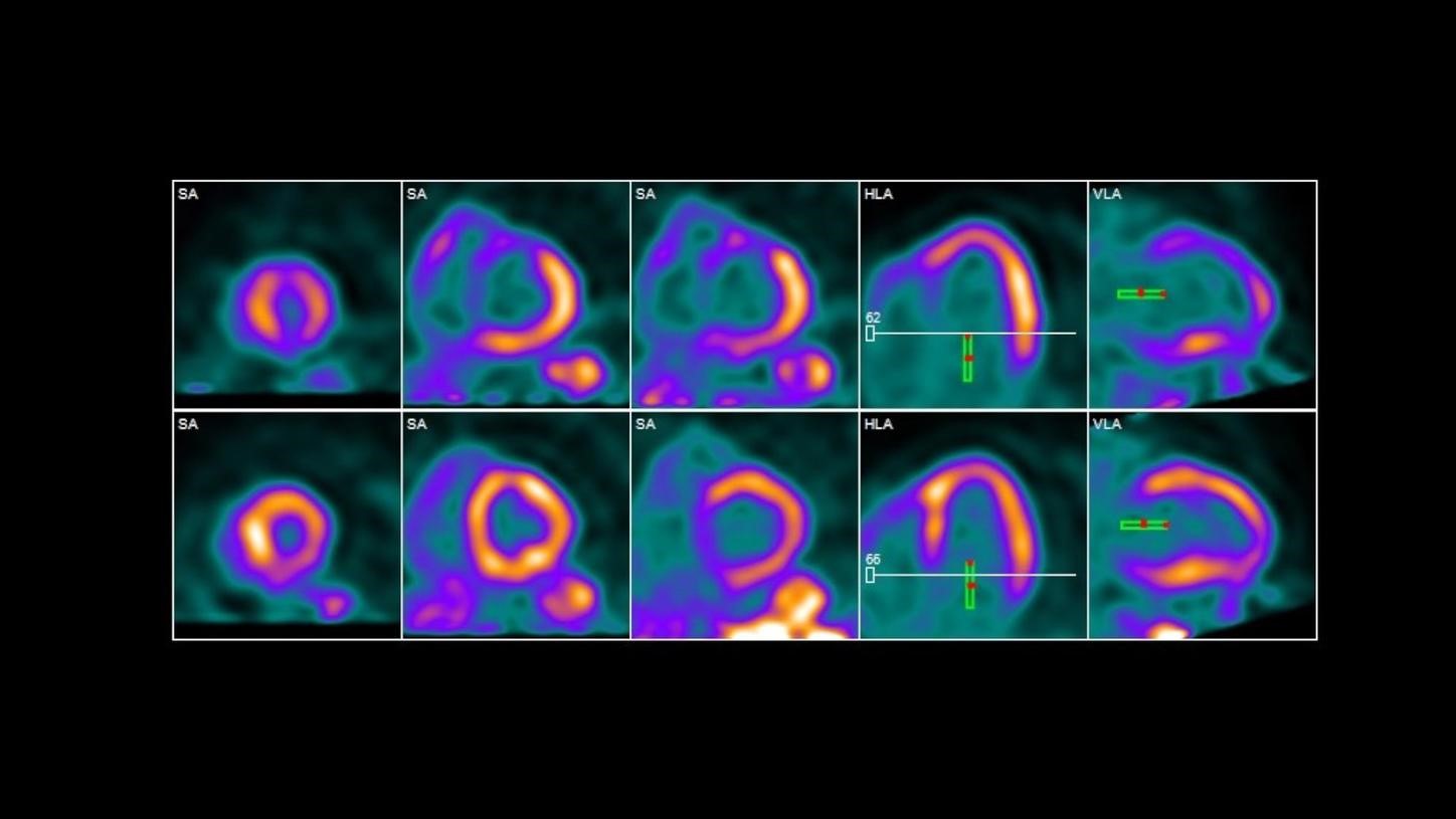
Comparison Phases
To get a full picture, the scan is typically done in two stages:
- Rest Phase: Images are taken while you are lying still to see normal blood flow.
- Stress Phase: You receive medication (like Lexiscan) that dilates your heart's arteries, simulating the effects of exercise. A second set of images is taken to see how blood flow changes under "stress".
3D Image Creation
A computer combines the data from thousands of these photon detections to build a detailed 3D map of your heart. This allows doctors to quantify blood flow in exact milliliters per minute, which is more precise than other standard imaging tests.
A cardiac PET scan is an advanced diagnostic tool used to evaluate complex heart conditions that standard tests—like EKGs or echocardiograms—might miss. It provides a detailed, functional look at the heart's blood supply and cellular health to guide critical treatment decisions.
Doctors typically order this scan for three main reasons:
- Coronary Artery Disease (CAD) Diagnosis: It is highly accurate at identifying narrowed or blocked arteries. It can detect early atherosclerosis (plaque buildup) and quantify absolute blood flow in milliliters per minute, which helps identify "balanced" blockages where all major vessels are narrowed.
- Myocardial Viability Testing: Before high-risk surgeries like bypass (CABG) or stenting, a PET scan determines if a damaged part of the heart is truly dead (scar tissue) or just "hibernating" due to low blood flow. If the tissue is viable, the surgery is more likely to improve heart function.
- Detecting Inflammation & Infection: It is a primary tool for diagnosing cardiac sarcoidosis, a rare inflammatory disease. It can also detect infections in heart valves or implanted devices like pacemakers.
A cardiac PET scan is often the preferred choice for specific patient groups:
- Inconclusive Results: Used when a standard stress test or SPECT scan provides unclear data.
- Body Habitus: Its higher energy tracers provide clearer images for patients with a high BMI or large amounts of chest tissue that might obstruct other scans.
- Risk Assessment: It helps predict future cardiac events (like heart attacks) by identifying microvascular disease—damage to the tiny vessels that are invisible on a standard angiogram.
A cardiac PET scan is considered the gold standard for noninvasive heart imaging because it provides detailed functional data that other tests cannot. Its primary benefits center on superior accuracy, lower radiation, and the ability to detect disease much earlier.
Superior Accuracy and Detail:
- Highest Diagnostic Precision: PET has higher sensitivity and specificity than standard stress tests or SPECT scans. It is better at detecting small blockages and subtle blood flow changes that might be missed elsewhere.
- Quantitative Measurements: Unlike tests that only provide relative visual comparisons, PET measures absolute blood flow in milliliters per minute. This helps identify "balanced" disease, where all major arteries are narrowed equally, a condition often missed by other scans.
- Tissue Viability: It is the most reliable way to distinguish between dead scar tissue and "hibernating" muscle that can be saved with surgery.
Patient Safety and Convenience:
- Lower Radiation: Cardiac PET typically uses 2 to 3 times less radiation than a traditional SPECT nuclear stress test.
- Faster Procedure: The entire scan often takes only 60 to 90 minutes, compared to the 3 to 4 hours required for some other nuclear imaging methods.
- No Physical Stress Required: Because it uses medication to simulate exercise, it is ideal for patients who cannot use a treadmill due to mobility or health issues.
Effectiveness for Diverse Body Types:
Traditional heart scans can be less accurate for certain patients due to "attenuation artifacts"—where body tissue blocks the signal. PET scans are not compromised by these factors, making them highly reliable for:
- Patients with a high BMI (obesity).
- Patients with large breasts or chest implants.
- Patients with fluid buildup around the lungs.
Strategic Treatment Planning:
- Avoids Unnecessary Surgery: By clearly identifying which areas of the heart will truly benefit from blood flow restoration, it helps doctors avoid invasive procedures that wouldn't help.
- Early Detection: It can identify the earliest stages of plaque development before symptoms even appear, allowing for earlier preventive care.
Before undergoing a cardiac PET scan, you must follow strict preparation guidelines to ensure the radioactive tracers can provide accurate images of your heart. Failure to follow these can result in the test being rescheduled.
Diet and Caffeine Restrictions:
- Zero Caffeine: You must avoid all caffeine for 12 hours before the test. This includes:
- Coffee and tea (even decaf, which contains trace amounts).
- Sodas and chocolate.
- Pain relievers containing caffeine, such as Excedrin or Anacin.
- Fasting: Most centers require you to fast for 4 hours prior, drinking only plain water. Avoid gum, mints, and cough drops.
- Special Diets: If checking for cardiac sarcoidosis or infection, you may be asked to follow a high-fat, low-carb diet for 24–48 hours beforehand.
Medication Management:
- Asthma and Heart Drugs: Certain medications, such as Theophylline or Aggrenox, must often be held for 48 hours as they can interfere with the stress agent.
- Beta-Blockers: Ask your doctor if you should stop these, as they can mask heart issues during the stress portion of the test.
Personal Conditions:
- Pregnancy/Nursing: Notify your provider immediately. Radiation can be harmful to a fetus, and nursing mothers may need to pump and store milk for 24 hours post-scan.
- Claustrophobia: The scanner is a donut-shaped tunnel. If you are anxious in small spaces, ask for a mild sedative beforehand; if you take one, you will need a driver to take you home.
- Physical Activity: Avoid strenuous exercise for 24 hours before the scan, as it can affect how the tracer is absorbed by your muscles instead of your heart.
Clothing and Metal:
- Wear loose, comfortable clothing.
- Avoid metal, including jewelry, zippers, underwire bras, and belts, as these can create shadows on the images.
Important Reminders:
- Consult your doctor: Always discuss any concerns about the risks or suitability of a PET scan with your doctor.
- Provide medical history: Be sure your doctor is aware of all your medical conditions, medications, and allergies.
- Follow instructions: Adhere to any instructions given by your healthcare provider regarding diet, medication, and preparations for the scan.